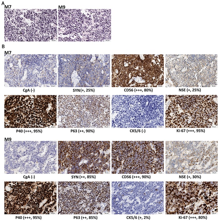
Figure 1. Histopathological diagnosis for the two MANECs.

(A) HE staining, showing cell features of small size, round or oval shape, hyperchromatic nuclei, and scanty cytoplasm. Nucleoli is inapparent and mitoses is prominent. (B) IHC staining. Neuroendocrine markers include CgA, SYN, CD56 and NSE. Squamous differentiation markers are P40, P63 and CK5/6. The marker for active proliferation is Ki-67. IHC staining demonstrates the coexistence of poorly differentiated neuroendocrine and squamous features in the same tumor. The expression levels are determined by the intensity of IHC staining. “-” indicates no stain. “+” indicates light yellow granulum and broken cell membrane. “++” indicates yellow granulum and intact cell membrane. “+++” indicates brown granulum, intact cell membrane and uniform staining. Mean positive staining percentages were calculated by averaging over ten randomly selected views for each sample.
