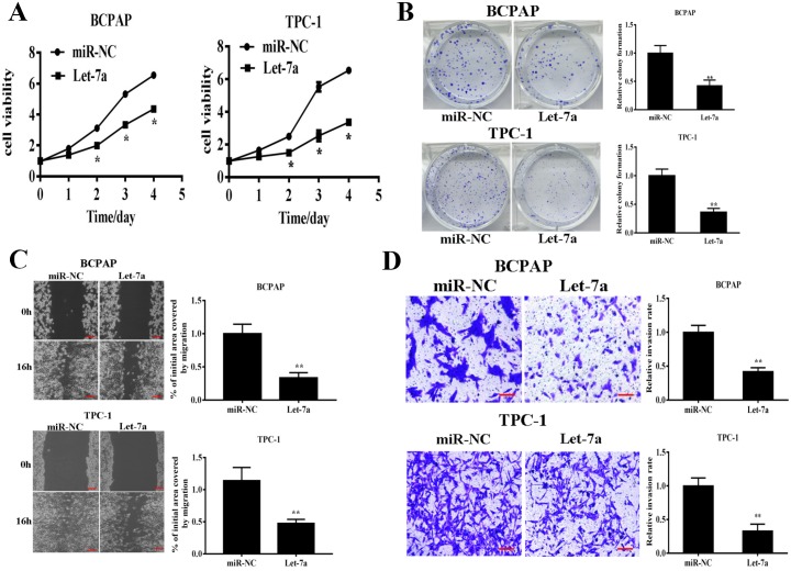
Figure 2. Let-7a overexpression inhibits cell proliferation, colony formation, migration and invasion.
BCPAP and TPC-1 cells were infected with lentivirus packing let-7a and expression. (A) Cell proliferation was assessed by CCK-8 kit. (B) Analysis of colony formation was shown. (C) Cells were cultured until reached 90% confluence. 20μl tips were used to scratch the cells layers to form a wound. The wound gaps were photographed and measured. (D) Transwell invasion assays was performed. After fixed, stained, and photographed, the cells in the bottom of the invasion chamber was measured by the absorbance at 570nm. Error bars represent the mean±SD of triplicate experiments. (**) Significant difference when compared with the miR-NC group (P< 0.01). Bar =200μm.

