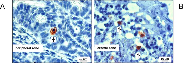
Figure 4. Typical M30 CytoDEATH stains of apoptotic cells, exemplarily shown for a mouse of the control cohort.
Apoptotic cells were very scarce in the periperal zone of the tumors (A), and somewhat more frequent (but still rare) in the transition zone to the necrotic centre of the xenografts (B). Arrows point to positive-stained cells.

