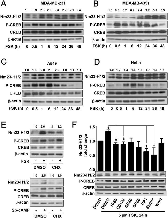
Figure 4. Time-dependent upregulation of Nm23-H1/2 protein expression in various cell lines.
MDA-MB-231 (A), MDA-MB-435s (B), A549 (C) and HeLa (D) cells were serum-starved overnight, followed by 5 μM forskolin treatment for different periods of time (0.5, 1, 6, 12, 24, 36, 48 h). Cell lysates were immunoblotted for detecting Nm23-H1/2, CREB and phosphorylated CREB. Numerical values shown above the immunoreactive bands represent relative intensities of Nm23-H1/2 as a ratio of the basal level at time 0 (set as 1.0). (E) HEK293 cells were pre-treated with 50 μM cycloheximide (CHX) in serum-free medium for 4 h and then treated with or without 5 μM forskolin or 10 μM Sp-cAMP overnight. (F) HEK293 cells were pre-treated with inhibitors specific for different signaling intermediates: 10 μM H-89 (PKA), 10 μM U0126 (ERK), 10 μM SB203580 (p38), 10 μM SP600125 (JNK), 10 μM PP1 (c-Src), 5 μM Stattic (STAT3), and 100 nM wortmannin (PI3K) for 4 h, followed with 5 μM forskolin treatment overnight. Cell lysates were subjected to Western blotting. *Forskolin significantly upregulated the protein expression of Nm23-H1/2 as compared to the vehicle control (P < 0.05); variations within control groups are <5%; ‡Significantly inhibited as compared to forskolin alone (P < 0.05).

