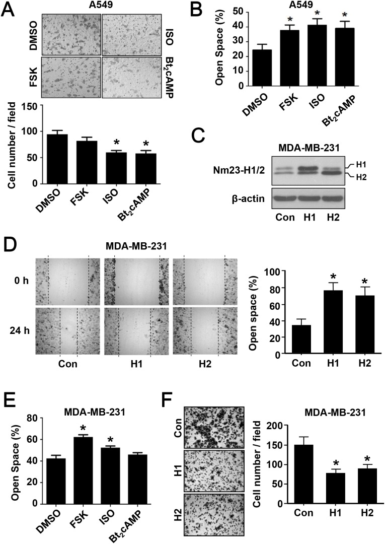
Figure 6. The effect of PKA activation on cancer cell migration.
(A) Metastatic lung cancer A549 cells were subjected to the following drug treatment for 24 h in serum-free medium: vehicle (DMSO, 0.5% v/v), forskolin (FSK, 5 μM), isoproterenol (ISO, 1 μM), and Bt2cAMP (10 μM). After the treatment, cells were reseeded into the inserts of transwell plates and allowed to migrate for 24 h prior to fixation and staining. Images were obtained with an Olympus 1X-HOS imaging system at 100× magnification and representative photos of each condition are shown. Quantification of the images is shown below the micrographs. (B) Confluent monolayer of A549 cells was scratched with a yellow tip and washed with PBS. Cells were allowed to migrate into the open wound for 24 h in the absence or presence of PKA activating agents as in panel A. Images were taken immediately after creating the wound and at 24 h. The open space (%) was the ratio of wounded area not covered by migratory cells after migration to that of time zero. (C) Metastatic breast cancer MDA-MB-231 cells stably overexpressing Nm23H1 or Nm23H2 were established as described in Materials and Methods. Cell lysates were analyzed by Western blotting with an anti-Nm23H1/2 antiserum. (D) MDA-MB-231 cells overexpressing Nm23H1 or Nm23H2 were subjected to the wound healing assay. (E) Parental MDA-MB-231 cells were treated with PKA activators and assayed as in panel B. (F) MDA-MB-231 cells overexpressing Nm23H1 or Nm23H2 were subjected to transwell migration assay as in panel A. *Significantly different to that of the corresponding vehicle or control group (P < 0.05).

