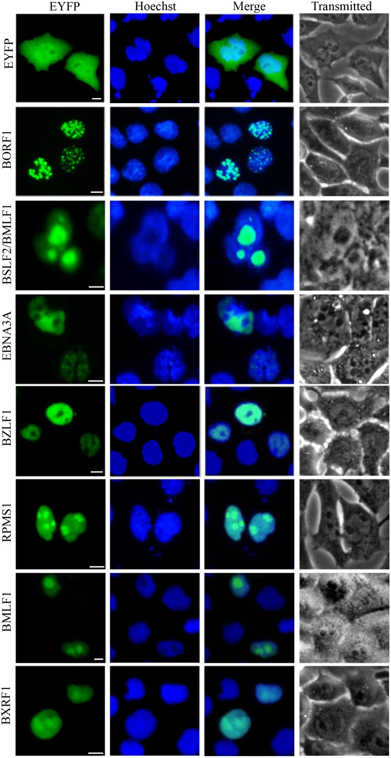
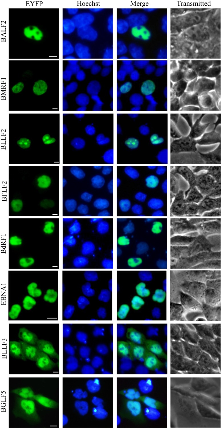
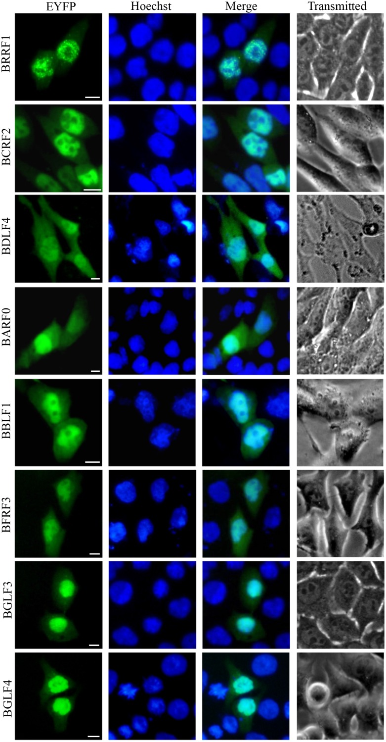
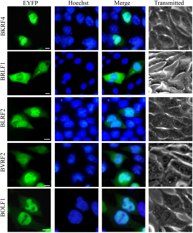
Figure 1. Nuclear localization summary of EBV-encoded proteins.
28 EYFP-fused EBV proteins were expressed in COS-7 cells, and cells were subjected to fluorescence microscope analysis in live cells 24 h after transfection. As a negative control, cells were transfected with the vector control (pEYFP-C1). Pictures were obtained using a Zeiss Axiovert 200M microscope. The same magnification was used in all panels. Representative fluorescence images of the vast majority live cells expressing indicated fusion protein were shown. Cells were counterstained with Hoechst to visualize the nuclear DNA. All scale bars indicate 10 μm.


