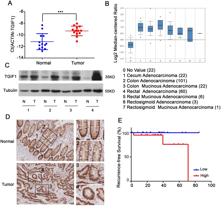
Figure 1. TGIF1 is highly expressed in CRC.
(A) qRT-PCR analysis of TGIF1 mRNA in CRC tissues (Tumor) and paired normal tissues (Normal). (B) TGIF1 expression analysis in all kinds of CRC according to Oncomine Database (https://www.oncomine.org/resource/login.html). (C-D) TGIF1 protein level in CRC tissues (T) and paired normal tissues (N) were assessed by immunoblotting (C) and immunohistochemistry (D) respectively. Enlargements in right: 1, stem cell region of normal crypts; 2, differentiation region of normal crypts; 3, 4, colon tumor regions. (E) Kaplan-Meier graph representing the probability of cumulative recurrence-free survival in CRC patients according to the TGIF expression in TCGA database, low (20) versus high (20). p=0.086.

