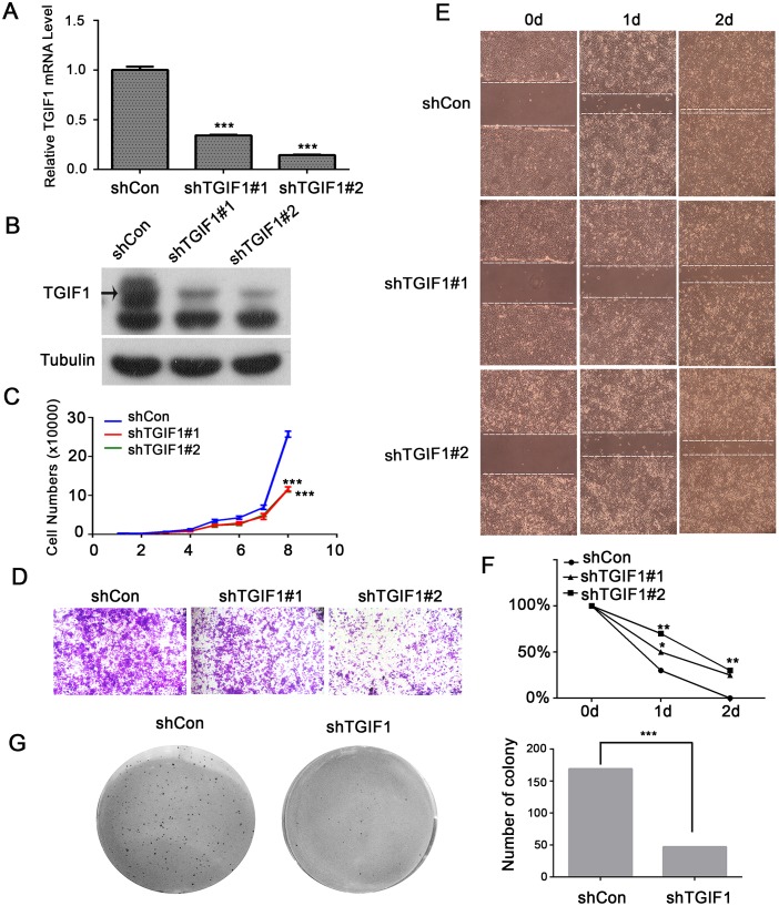
Figure 2. TGIF1 enhances proliferation, migration and clonogenicity of LoVo cells.
(A-B) qRT-PCR and immunoblotting showing TGIF1 knockdown efficiency in LoVo cells. (C) Growth curve of control and TGIF1 knockdown cells. The data are presented as the mean ± S.E. ***, p < 0.001. (D) Transwell assay using control and TGIF1 knockdown cells. After seeding cells for 24 hours, the number of migrated cells was quantified after staining with gentian violet. (E-F) Wound healing assay in control and TGIF1 knockdown cells. Cells were photographed every 24h after scratching. Statistical results were shown by linear graph. *, p<0.05;**, p<0.01. (G) Soft agar colony formation assay using control and TGIF1 knockdown cells. Images were obtained after staining with Giemsa.

