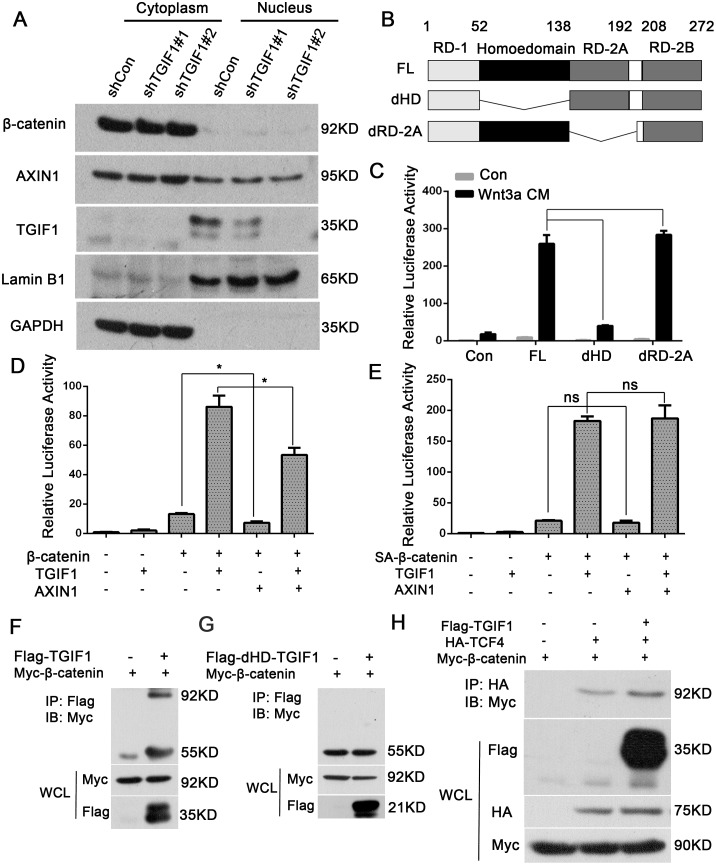
Figure 5. The TGIF1 homeodomain is essential for promoting Wnt signaling.
(A) Nuclear and cytoplasmic fractions were separated from LoVo cells and subjected to immunoblotting with the indicated antibodies. (B) Diagrams showing TGIF1 domain deletion constructs. FL for full length; dHD for homeodomain deletion and dRD-2A for RD-2A domain deletion. (C) Wild-type TGIF1 and various deletion constructs were co-transfected with Topflash-luciferase into HEK293T cells. After 24 hours, the cells were treated with Wnt3a conditional medium for another 24 hours and then harvested to measure luciferase activity. ***, p < 0.001; ns, p>0.05. (D-E) HEK293T cells were transfected with Topflash-luciferase together with TGIF1, AXIN1, wild-type β-catenin or SA-β-catenin as indicated. At 48 hour post-transfection, the cells were harvested for luciferase assay. *, p <0.05; ns, p>0.05. (F-G) HEK293FT cells were co-transfected with constructs carrying β-catenin, TGIF1 and dHD-TGIF1 as indicated for 48h before they were harvested. Cell lyses were precipitated with anti-Flag antibody, followed by anti-Myc immunoblotting (IB) to detect the interaction between β-catenin and TGIF1. IP, immunoprecipitation; WCL, whole cell lysate. (H) HEK293FT cells were co-transfected with constructs carrying β-catenin, TGIF1 and TCF4 as indicated for 48h before they were harvested. Cell lyses were precipitated with anti-HA antibody, followed by anti-Myc immunoblotting (IB) to detect the interaction between β-catenin and TCF4.

