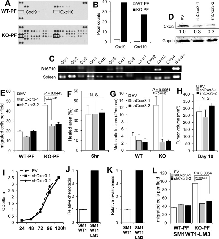
Figure 3. Melanoma-encoded Cxcr3 is required for the enhanced peritoneal metastasis in KO mice and for the enhanced chemotaxis to KO-PF.
(A) Antibody array blots of equal protein aliquots of PF from WT- or KO-mice, identifying the paired spots for Cxcl9 and 10 (boxes). (B) Quantification of the pixel counts in the blot in panel A. (C) Semi-quantitative RT-PCR for various chemokine receptors in B16F10 cells, using total spleen cells from WT mice as a control. The primer sets used are described previously [62]. (D) IB of B16F10-luc stably expressing empty vector (EV) or clones of Cxcr3-specific shRNA, blotted for either Cxcr3 or Gapdh. (E) Chemotaxis assay of B16F10-luc cells shown in panel 2D towards media containing 20% PF from WT- or KO-mice. (F) Percent healed area of wound monolayers from the cells in panel E in media containing 20% KO-PF. (G) Number of peritoneal metastases/mouse after the i.v. injection of B16F10-luc cells (EV, shCxcr3-1 or shCxcr3-2) into WT- or KO hosts (n = 12 each). (H) Tumor volumes (s.c.) of B16F10-luc[EV], B16F10-luc[shCxcr3-1] or B16F10-luc[shCxcr3-2] cells in KO mice (n = 6). (I) Relative cell numbers of B16F10-luc[EV], B16F10-luc[shCxcr3-1] or B16F10-luc[shCxcr3-2] cells. (J) Relative chemotaxis of SM1WT1-luc or SM1WT1-LM3-luc cells towards media containing 10% FBS. P < 0.0001. (K) Relative Matrigel invasiveness of SM1WT1-luc or SM1WT1-LM3-luc cells. P < 0.0001. (L) Chemotaxis assay towards media containing 20% PF from WT- or KO-mice by SM1WT1-LM3-luc cells transduced with EV, shCxcr3-1 or -2.

