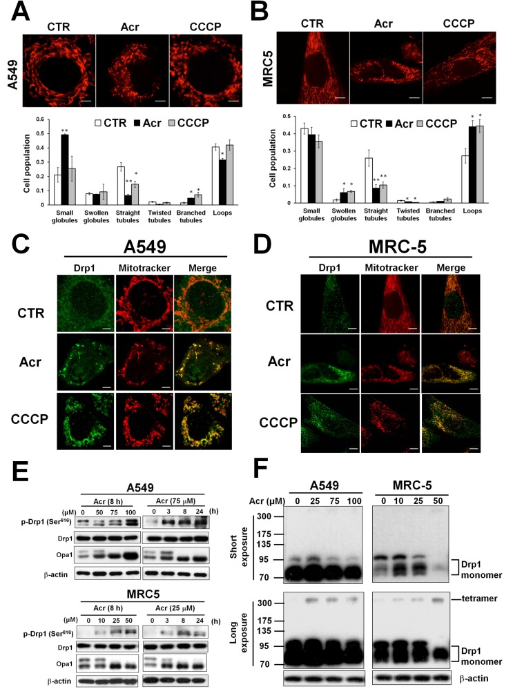
Figure 6. Acrolein induces mitochondrial fission.
In panel (A & B), A549 and MRC-5 cells were treated with Acr (A549: 75 μM, MRC-5: 25 μM) for 8 h, and stained with mitotracker (upper) and the mitochondrial morphology change was quantified (lower). Bar graphs show data collected from 3 independent experiments. Data are mean ± s.d. * P< 0.05; ** P<0.01. In panels (C & D), Drp1 translocation was detected by immunofluorescent staining assay. A549 and MRC-5 cells were treated with Acr (A549: 75 μM, MRC-5: 25 μM) or CCCP (20 μM) for 8 h at 37°C, fixed, stained with Drp1, and then examined by microscopy. Mitotracker was used to stain mitochondria. Phosphorylated Drp1 (p-Drp1, Ser616), Drp1, OPA1 (panel E) and tetramerization of Drp1 (panel F) in Acr-treated cells was detected by Western blot analysis. Note: Acr induces phosphorylation of Drp1 at Ser616 site followed by tetrameriaztion of Drp1 and cleavage of OPA1 indicating that Acr induces mitochondrial fission.

