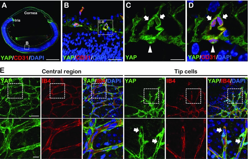
Fig. S1.
YAP expression in the developing retinal vasculature. (A–D) YAP staining images of cross-section of P5 wild-type eyes. High magnification images (C and D) showed extranuclear (arrows) and nuclear (arrowheads) YAP localization in retina VECs. (E) Whole-mount YAP staining of P5 wild-type retina. The higher magnification of the boxed areas (Upper) are shown (Lower). Tip cells showed extranuclear YAP localization (arrows). [Scale bars: 500 μm (A), 200 μm (E, Upper), 50 μm (B and E, Lower), and 12.5 μm (C).]

