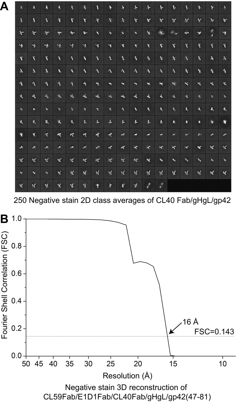
Fig. S5.
Negative-stain EM of gHgL/gp42 with different Fab complexes. (A) A total of 250 2D class averages for CL40Fab/gHgL/gp42 showing a general T-shaped structure with the CL40Fab bisecting the rod-shaped gHgL. This model closely aligns with the crystal structure of CL40Fab/gHgL/gp42(47–81). Here the gp42 CTLD or C-domain is displaced due to the presence of CL40Fab sharing the same gH-binding site. This is seen in certain 2D classes, in cases of gp42 density overlapping or to the side of gHgL, and with CL40 bound as shown in Fig. 4D. (Scale: panels are 35 × 35 nm.) (B) FSC vs. resolution curve after 3D postrefinement for the CL59Fab/E1D1Fab/CL40Fab/gHgL/gp42(47–81) complex.

