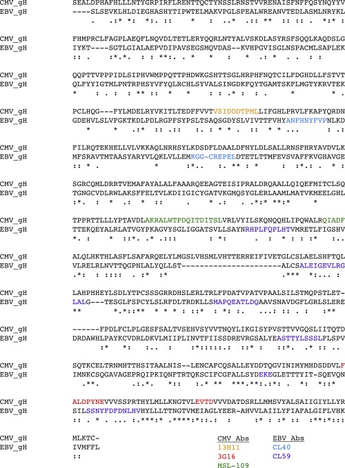
Fig. S6.
CMV- and EBV-gH–neutralizing antibody epitopes. Sequence alignments of the ectodomains of gH from CMV and EBV with corresponding antibody epitopes are highlighted. The antibody epitopes are derived by different methods, including crystal structure, negative-stain EM, and hydrogen-deuterium exchange mass spectrometry (HDX-MS) by Ciferri et al. (45, 46). The linear epitopes of CL40 (blue) and 13H11 (orange) are in a similar region, and epitopes of 3G16 (red), MSL-109 (green), and CL59 (purple) correspond to D-IV of gH. Together with previous site-directed mutagenesis that have a cell-type–specific fusion defect, these Ab epitopes highlight common regions of vulnerability in gHgL across different herpesvirus subfamilies.

