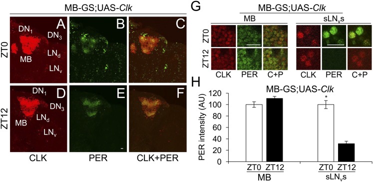
Fig. 6.
Clk expression in MB neurons does not support PER cycling in LD. MB-GS/+; UAS-Clk/+ (MB-GS; UAS-Clk) flies were induced with RU486, entrained and collected as described in Fig. 4. Immunostaining with CLK and PER antibodies was performed on dissected adult brains and imaged by confocal microscopy. Projected Z-series images of right brain hemispheres are shown, where lateral is right and dorsal is top. Pacemaker neuron groups are as defined in the text, and MB neurons are as defined in Fig. S1. Colocalization of CLK (red) and PER (green) is shown as yellow. (A–C) A 136-μm projected Z-series image of a brain from flies collected at ZT0 and immunostained with CLK (A), PER (B), or CLK and PER (C). (D–F) A 136-μm projected Z-series image of a brain from flies collected at ZT12 and immunostained with CLK (D), PER (E), or CLK and PER (F). (Scale bar, 10 μm). Panels A–F are the same magnification. (G) Magnified 2-μm images of MB neurons (Left) and magnified 18-μm projected Z-series images of sLNvs (Right) from flies collected at ZT0 in A–C or at ZT12 in D–F. (Scale bar, 10 μm.) C+P, CLK + PER. All images are representative of six or more brains. (H) PER immunostaining intensity was quantified in MB neurons and sLNvs from flies collected at ZT0 and ZT12. AU, arbitrary units. Error bars indicate ±SEM. PER intensity was significantly (*P < 0.01) higher in sLNvs at ZT0 than at ZT12 by two-tailed Student’s t test.

