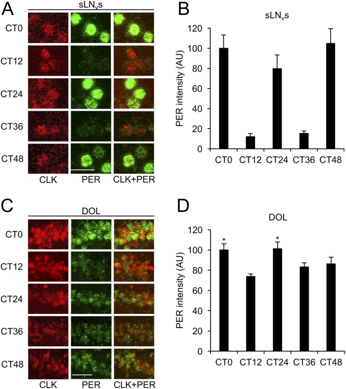
Fig. S2.
Clk expression in DOL cells supports PER cycling that rapidly dampens in DD. The 3.0cry-Gal4/+; UAS-Clk/+ flies were entrained in LD cycles for 3 d, transferred to constant darkness, and collected at CT0, CT12, CT24, CT36, and CT48. Immunostaining with anti-PER and anti-CLK antibodies was performed on adult brains and imaged by confocal microscopy. Colocalization of CLK (red) and PER (green) is shown as yellow. (A) The 22-μm projected Z-series images of sLNvs from flies collected at the indicated times and immunostained with CLK (Left column), PER (Middle column), or CLK and PER (Right column). (B) PER immunostaining intensity from sLNvs was quantified in AU as described in Materials and Methods. Error bars indicate ±SEM. The overall effects of time of day were significant (P < 0.0001) by one-way ANOVA. Time-dependent cycling was significant (P < 0.05) by Tukey post hoc analysis. (C) The 26-μm projected Z-series images of DOL cells from flies collected at the indicated times and immunostained with CLK (Left column), PER (Middle column), or CLK and PER (Right column). (D) PER immunostaining intensity from DOL cells was quantified in AU as described above. Error bars indicate ±SEM. The overall effects of time of day were significant (P < 0.01) by one-way ANOVA. Time-dependent cycling was not significant by Tukey post hoc analysis. Asterisks denote significant (P < 0.05) increase in PER in DOL cells at CT0 and CT24 compared with CT12 by two-tailed Student’s t test. (Scale bars, 10 μm.) All images in panels A and C are the same magnification. All images are representative of six or more brains.

