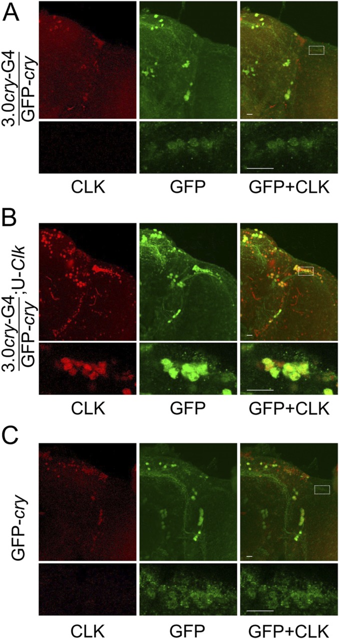
Fig. S3.
Endogenous CRY expression in DOL cells is enhanced by Clk expression. The 3.0cry-Gal4/GFP-cry (3.0cry-G4/GFP-cry), 3.0cry-Gal4/GFP-cry;UAS-Clk/+ (3.0cry-G4/GFP-cry;U-Clk), and GFP-cry flies were entrained in LD cycles for 3 d, transferred to constant darkness, and collected at CT24 on the first day of DD. Immunostaining with CLK and GFP (to detect GFP-CRY) antibodies was performed on adult brains and imaged by confocal microscopy. Colocalization of CLK (red) and GFP (green) is shown as yellow. (A) The 130-μm projected Z-series images of the right hemisphere from a 3.0cry-G4/GFP-cry brain (Top) and magnified 2-μm sections of the DOL region (Bottom) from the brain on Top immunostained with CLK (Left column), GFP (Middle column), or CLK and GFP (Right column). White rectangle, DOL region used for magnified image on Bottom. (B) The 140-μm projected Z-series images of the right hemisphere from a 3.0cry-G4/GFP-cry;U-Clk brain (Top) and magnified 2-μm sections of the DOL region (Bottom) from the brain on Top immunostained with CLK (Left column), GFP (Middle column), or CLK and GFP (Right column). White rectangle, DOL region used for magnified image on Bottom. (C) The 146-μm projected Z-series images of the right hemisphere from a GFP-cry brain (Top) and magnified 2-μm sections of the DOL region (Bottom) from the brain on Top immunostained with CLK (Left column), GFP (Middle column), or CLK and GFP (Right column). White rectangle, DOL region used for magnified image on Bottom. All images are representative of six or more brains. (Scale bars, 10 μm.) All top images and all bottom images in panels A–C are the same magnification.

