Fig. 1.
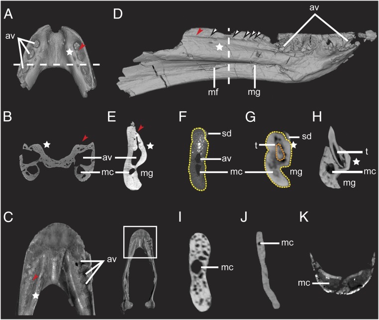
Dinosaurian and crocodilian dentaries illustrated by CT data, showing the presence of the alveolar canal and other alveolar vestiges. (A) Dorsal view and (B) coronal section of Caenagnathasia sp. (IVPP V20377). (C) Dorsal view of rostral dentary of cf. Chirostenotes TMP 2012.12.12, and (Inset) entire mandible. Photographs in Fig. 1C are reproduced with permission from ref. 24. Copyright 2008 Canadian Science Publishing or its licensors. (D) Mirror symmetry of right dentary of Sapeornis chaoyangensis (LPM B00015) in lingual view showing vestigial alveoli and foramina present on the dorsolingual aspect of the dentary. (E–K) Coronal dentary sections of (E) S. chaoyangensis (LPM B00015), (F) subadult Limusaurus inextricabilis (IVPP V15923), (G) juvenile L. inextricabilis (IVPPV15301), (H) extant Alligator sinensis (IVPP 1361), (I) extant Pavo sp. (IVPP 1032), (J) Confuciusornis sp. (IVPP V23275), and (K) Khaan mckennai (IGM 100/973). av, alveolar vestige; mc, mandibular canal (V3); mf, Meckelian foramen; mg, Meckelian groove; sd, supradentary; t, tooth. White stars, swollen ridge present on the lingual aspect of the dentary; red arrows, lingual groove; white arrows, foramina piercing the lingual groove on the medial aspect of the dentary; dashed lines in A and D mark the positions of the slices shown in B and E, respectively; light yellow dashed lines in F and G mark the dentary; orange dashed line in G marks a replacement tooth present inside an enclosed dentary alveolus. (Not to scale.)

