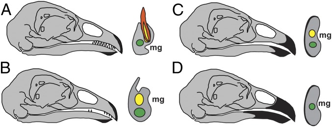
Fig. 4.
Transformations involved in theropod tooth reduction. Right lateral view of the head (Left) and transverse view of the dentary (Right). (A) Normal tooth development and tooth replacement with apomorphic keratinized rhamphotheca covering only the rostral-most portion of the jaws. (B) Tooth replacement is impeded by external closure and/or constriction of alveoli and regional tooth reduction occurs. (C) As the keratinized rhamphotheca enlarges, the remaining teeth are either functionally reduced or redundant. (D) Edentulous beak completely covered by rhamphotheca. Green, mandibular canal; yellow, alveolus and alveolar canal; orange, tooth. (Not to scale)

