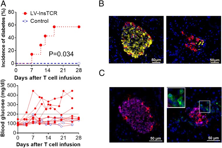
Fig. 4.
Induction of diabetes in HLA-DQ8–Tg hu-mice by adoptive transfer of autologous diabetogenic human CD4+ T cells. HLA-DQ8–Tg hu-mice were treated with low-dose streptozotocin and injected 1–2 d later with 5 × 10⁶ expanded LV-insTCR–transduced or control (i.e., the same hu-mouse–derived human CD4+ T cells that were similarly expanded in vitro as the LV-insTCR–transduced CD4+ T cells) human CD4+ T cells, followed 1 d later by immunization with InsB:9–23 peptides. (A) Cumulative incidence of diabetes (Top) and levels of blood glucose (Bottom) in hu-mice receiving LV-insTCR–transduced (solid symbol; n = 7) or control (opened symbol; n = 6) human T cells. Mice were defined as hyperglycemia if two consecutive blood glucose measurements >200 mg/dL (B and C) Immunofluorescent staining of pancreas samples prepared between 3 and 4 wk after injection of CD4+ T cells from hu-mice receiving control (Left) or LV-InsTCR–transduced (Right) human T cells (n = 3 per group). (B) Staining of mouse insulin (yellow) and glucagon (red). (C) Staining of human CD3+ cells (green), mouse insulin (pink) and glucagon (red). Nuclear is stained blue by DAPI.

