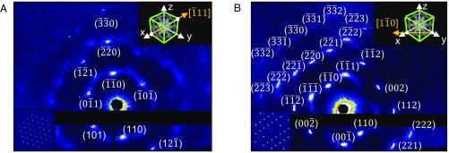
Fig. 4.
Single-crystal diffraction: 2D SAXS scans obtained from 6-wk-old samples (GMO/DOTAP/GMOPEG 97.5/1.5/1). (A) Single crystal aligned with direct beam at [−1 1 1] direction showing nine sharp Bragg spots. A simulated scattering pattern (Bottom Left Inset) is well matched with the data. (B) Single crystal aligned with direct beam at [1 −1 0] direction displaying 24 intense Bragg peaks. (Bottom Left Inset) A simulated scattering pattern is also perfectly matched with the data. The directions of the direct X-ray beam are shown by the yellow arrows in the Top Right Insets of (A) and (B).

