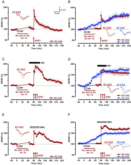
Fig. 3.
SP initiates STC in SC-CA2 and EC-CA2 synapses. (A) Experiment showing a WTET-induced early LTP in EC-CA2 synaptic inputs. After recording a stable baseline of 30 min, SP was bath-applied for 15 min, during which time the baseline stimulation was suspended for 1 h (n = 7). After that, a baseline was recorded for another 30 min followed by WTET protocol. The potentiation decayed to baseline level within 180 min. (B) STC initiated by SP. Stimulation of EC-CA2 inputs was suspended during the application of SP and, at the 90th minute, WTET was delivered and the SC-CA2 fEPSPs were further recorded. Unlike in A, SP transformed the EC-CA2 fEPSP potentiation into an L-LTP (n = 8). (C and E) Experiments similar to A but with STET protocol was applied at the 90th minute in the presence of protein synthesis inhibitors ANI (25 µM; n = 8; C) or EME (20 µM; n = 8; E). The inhibitors were applied 20 min before STET (i.e., 10 min after resuming the baseline recording) and washed out 40 min after STET of the EC-CA2 input. (D and F) STC experiments in which L-LTP in the EC-CA2 inputs (red circles) was expressed in the presence of protein synthesis inhibitors ANI (25 µM; n = 8; D) or EME (20 µM; n = 9; F) as a result of the capture of previously SP-induced plasticity-relevant proteins. The experimental design was similar to that in B with the exception that, in this case, STET was applied to EC-CA2 inputs at the 90th minute. Representative fEPSP traces 15 min before (closed line), 95 min after (dotted line), and 180 min after (hatched line) SP application or WTET/STET are depicted. Calibration bars for fEPSP traces are 2 mV/3 ms. Arrows indicate the time points of WTET or STET.

