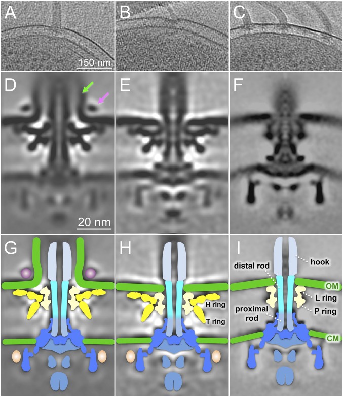
Fig. 1.
In situ V. alginolyticus flagellar structures: sheathed and unsheathed. (A) A representative slice of a 3D reconstruction of a wild-type V. alginolyticus VIO5 shows a single sheathed flagellum at the cell pole. (B) Another slice shows a single unsheathed flagellum. (C) A tomographic slice of the strain (KK148) shows multiple polar flagella. (D) A class average shows the sheathed flagellar motor structure. (E) Another class average shows the unsheathed flagellar motor structure. (F) An averaged structure of the E. coli motor is used as a reference for comparison. (G–I) Schematic models are overlaid on cryo-ET density maps of D–F, respectively. The key differences between Vibrio and E. coli motor structures are highlighted, with the O ring in purple, the H ring and T ring in yellow, and the extra density (colored in pink) located outside the C ring. CM, cytoplasmic membrane; OM, outer membrane. The stator density is not visible in the globally averaged structures of the Vibrio and E. coli motors likely because of the highly dynamic nature of the stators.

