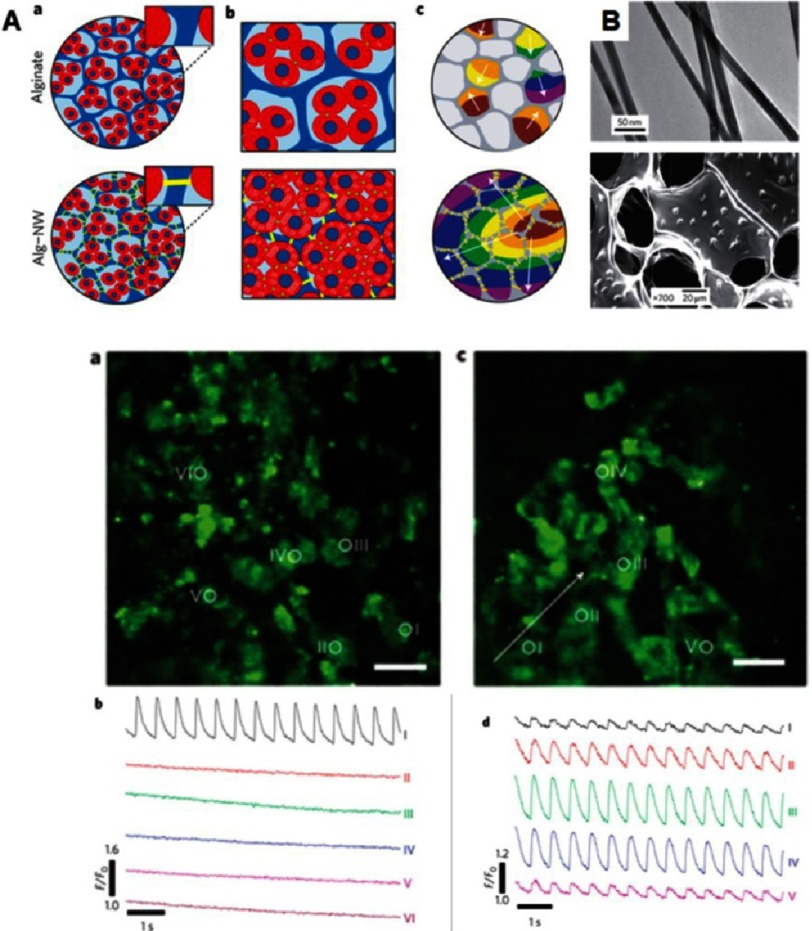
Figure 10. Cardiac tissue engineering in nano-composite multifunctional alginate scaffolds.
Schematic overview of three-dimensional engineered nano-wired cardiac tissue: alginate pore walls (blue), cardiac cells (red) and gold nanowires (yellow) (A). Incorporation of nanowires within alginate scaffolds (B). Top: typical gold nanowires, bottom: nanowires assembled within the pore walls of the scaffold. Cardiomyocytes cultured in pristine alginate (a) and in alginate nanowires–composites (c). Monitoring calcium dye fluorescence for measuring calcium transient propagation within engineered tissues assessed at points marked with white circles. Calcium transients were only observed at the stimulation point in the unmodified scaffold (b). Calcium transients were observed at all points of wired composite (d). Reprinted with permission from [78].

