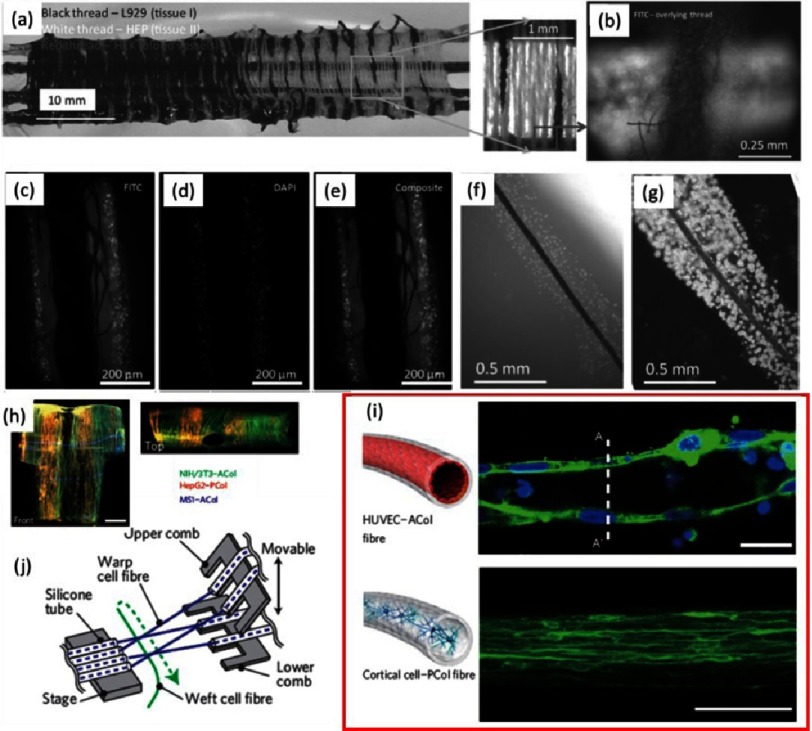
Figure 14. Alternative ways of 3D construct fabrication using alginate fibers.
Organ weaving: connecting tissues using gradients (A). An example of manual plain weaving using three cell lines. Magnified box: basic criss-cross pattern of plain weaving. Overlaid thread containing HepG2 cells and underlying thread containing HEK-293 cells labeled CellTracker Green CMFDA (B). Fiber-supported living threads, fluorescence images of double-treated thread, including fluorescently labeled L929 cells stained with CellTracker Green CMFDA (C) and MCF-7 cells stained with nuclei stain Hoechst-33342 (D). Overlay of images (C) and (D) showing two distinguishable layers of cells (E). Living thread immediately after preparation (F). The same after 8 days of incubation (G). Pictures were reprinted with permission from (10). Fiber-based assembly of higher-order 3D macroscopic cellular structures (H). Fluorescence micrograph of a HUVEC/ACol (top panel) and primary cortical cell/PCol fiber (bottom panel) at day 35 (I). Scale bars are 20 µm. Schematic of a microfluidic weaving machine working in culture medium (J). Pictures were reprinted with permission from [11].

