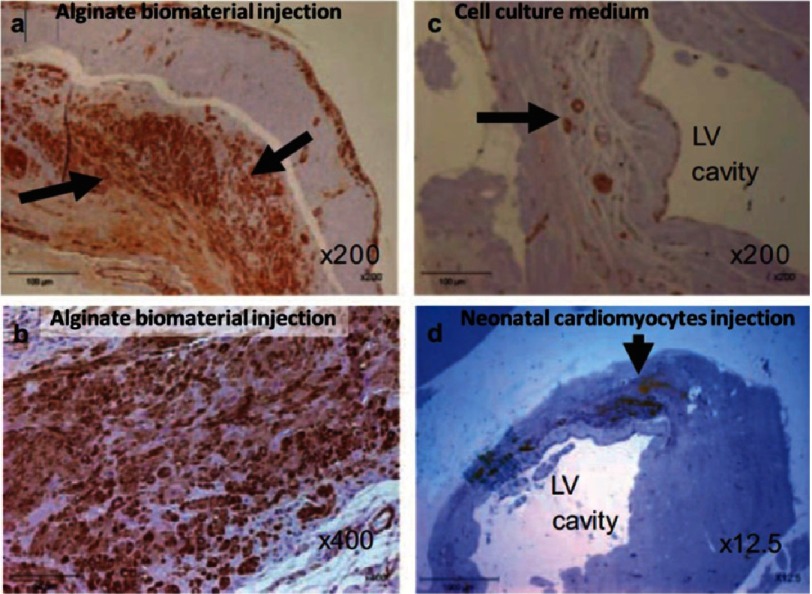
Figure 5. Infarcted rat hearts after immunostaining for α-SMA, after treatment with alginate or cardiomyocyte transplantation.
In the scar tissue 8 weeks after alginate injection, extensive positive brown staining revealed that the scar was populated with α-SMA positive myofibroblasts (A,B). Fewer myofibroblasts were observed for control scar to which the cell culture medium was injected in place of alginate (C). Neonatal cardiac cell implant (arrow) at the border of the infarct zone 8 weeks after transplantation (D). Engrafted cells were undifferentiated and isolated from the host myocardium. Reprinted with permission from [53].

