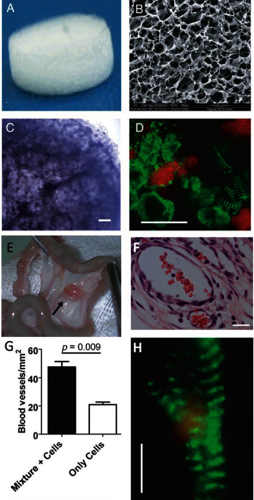
Figure 8. Construction of a cardiac patch in an alginate/alginate-sulfate scaffold capable of binding and releasing mixed factors.
The scaffold features before cell seeding; macroscopic view (A) and internal porosity by scanning electron microscopy (B). Cardiac patch seeded with cells and supplemented with factors after 48 h of cultivation. Light microscope view of the cardiac patch showing uniform distribution of cells in the matrix pores (C). Cardiac cell organization within the scaffold as judged by anti-actinin immunostaining (green) and nuclear staining (red) (D). Some of the cells reveal the typical striation of cardiac tissue. Vascularization of the 7 day omentum-transplanted mixture supplemented cardiac patch. The cardiac patch (arrow) is stitched to the omentum (E). Mature blood vessels populate the cardiac patch supplemented with factor as judged by anti-SMA immunostaining (brown) (F). Blood vessel density in the omentum-implanted patches (G). Typical cardiac cell striation is revealed in an omentum generated, factor-supplemented alginate cardiac patch as revealed by anti-actinin immunostaining (green) and confocal microscopy (H). Scale bar: 200 µm (C&E); 10 µm (D&H). Reprinted with permission from [87].

