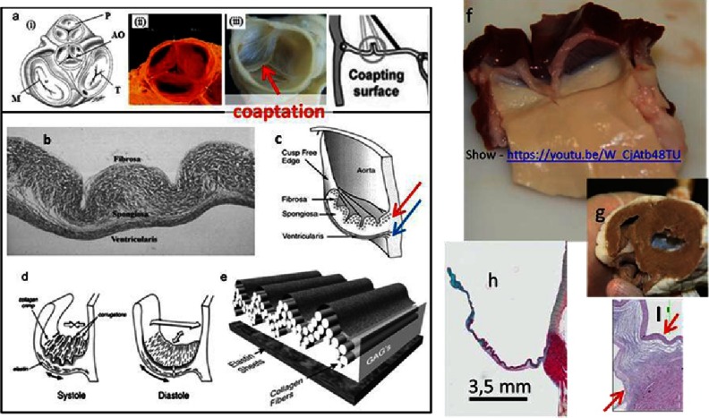
Figure 1. The basic structural/functional aspects of the heart valve (HV).
Schematics of the 2D position of the four valves on valvular basal plane of the heart where P: pulmonary valve, AO: aortic valve, M: mitral valve, and T: tricuspid valve (a-i), porcine pulmonary heart valve (a-ii), decellularized porcine aortic heart valve (a-iii). The leaflet consists of three distinguishable layers: fibrosa, spongiosa and ventricularis (b). Each layer contains specifically oriented fibers, red arrow indicates circumferentially oriented fibers of collagen, blue arrow indicates radially oriented fibers of elastin (c).The types (collagen, fibronectin, elastin and others) and arrangement of fibers are responsible for the function of the valve (opening & closing) (d, e). The fibrosa layer is a continuation of aorta and ventricularis is a continuation of the ventricular chamber. (f) The view of sheep aortic valve from ventricular side, after dissecting the heart (g). The leaflets contain glycosaminoglycans (GAGs, mainly in spongiosa, blue color), which works as a lubricant between layers and a shock absorber (h). The hinge area (in-between the arrows) histology highlights the importance of fiber arrangement (I). The cells are housed and maintained in a highly organized network of fibers. Reprinted with permission from4.

