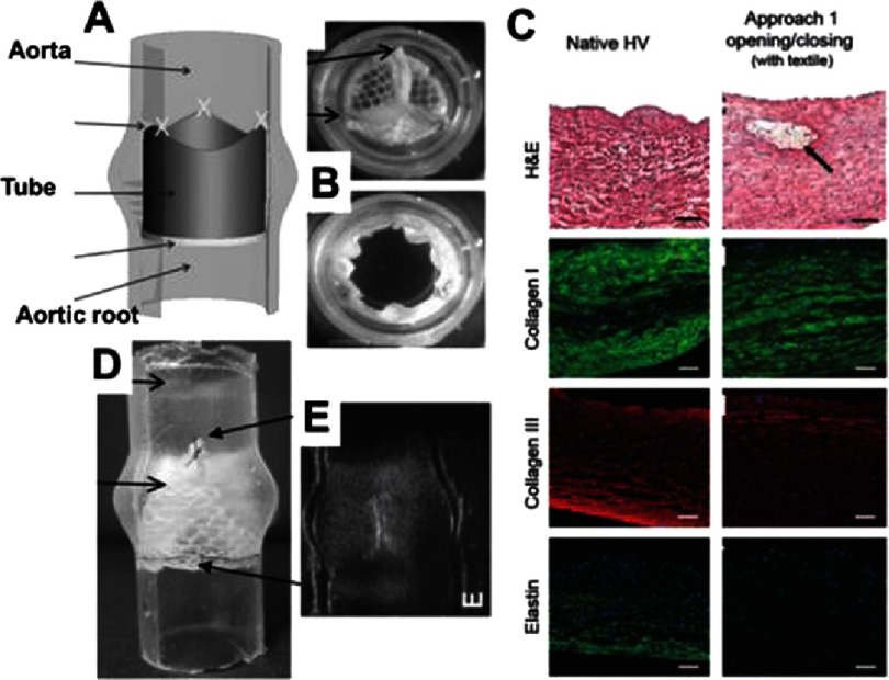
Figure 16. Tube-in-tube valve and its biocompatibility test.
Principle of the tube-in-tube valve (A). Still images of closed and open cycles of tube-in-tube valves (B). Histological and fluorescence immunohistochemical micrographs of native ovine pulmonary valve (wall), dynamically conditioned tissue-engineered tubular valves (C). Scale bars are 100 µm. The black arrow indicates the textile structure. Sutured in a silicone tube featuring the sinuses of Valsalva (D). Ultrasound images of the tube-in-tube in closed position (E). Reprinted with permission from51.

