
Figure 3. Lack of epithelial PPAR-γ-signaling increases colonocyte oxygenation during colitis.
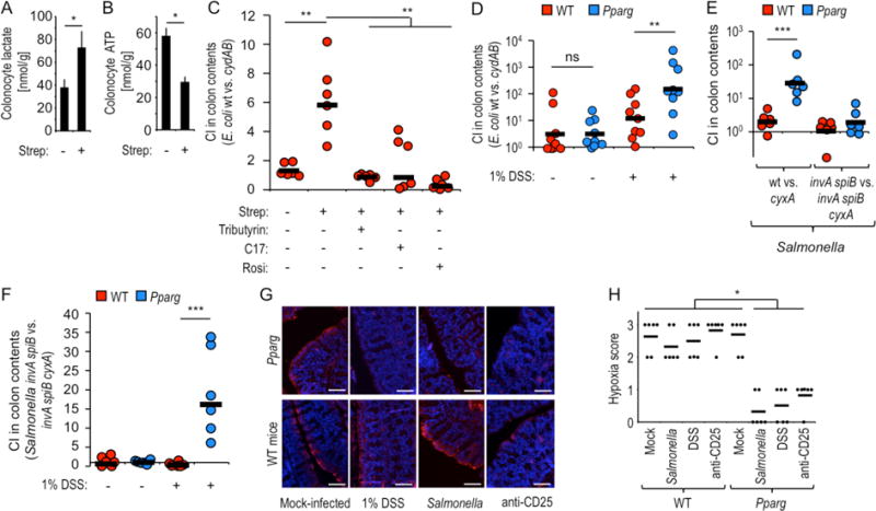
(A and B) Groups of mice (N = 5) were mock treated or treated with streptomycin (Strep) and colonocytes isolated one day later to measure intracellular concentrations of lactate (A) or ATP (B). (C) Groups (N = 6) of streptomycin-treated or mock-treated mice were inoculated with a 1:1 mixture of E. coli wild type (wt) and cydAB mutant and received supplementation with rosiglitazone (Rosi), tributyrin or a community of 17 human Clostridia isolates (C17). (D) Groups of mice (N = 6) receiving no supplementation or water supplemented with 1% dextran sulfate sodium (DSS) were inoculated with a 1:1 mixture of E. coli wild type (wt) and cydAB mutant. (C and D) The competitive index (CI) was determined 3 days after inoculation. (E and F) Groups of mice (N = 6) were inoculated with the indicated Salmonella strain mixtures. (F) Mice received no supplementation or water supplemented with 1% DSS. (G and H) Mice (N = 6) were treated as indicated and were injected intraperitoneally with pimonidazole one hour before euthanasia. Binding of pimonidazole was detected using hypoxyprobe-1 primary antibody and a Cy-3 conjugated goat anti-mouse secondary antibody (red fluorescence) in sections of the colon that were counter stained with DAPI nuclear stain (blue fluorescence). (G) Representative images are shown. Scale bars represent 50 μm. (H) A veterinary pathologist scored blinded sections for hypoxia staining. Each dot represents data from one animal. (A-B) Bars represent geometric means ± standard error. (C–F) Each dot represents data from an individual animal and black bars represent geometric means. *, P < 0.05; **, P < 0.01; ***, P < 0.001; ns, not statistically significantly different.