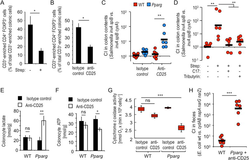
Figure 4. Microbiota-induced PPAR-γ-signaling and Tregs cooperate to limit the bioavailability of oxygen in the colon.

(A and B) Groups of mice (N = 4) were treated with streptomycin (A) or with anti-CD25 antibody (B) and CD3+-enriched live colonic cells analyzed for expression of CD4 and FOXP3 by flow cytometry. (C) Groups of mice (N = 6) were treated with anti-CD25 antibody or isotype control and 10 days later inoculated with a 1:1 mixture of an avirulent Salmonella strain (invA spiB mutant) and an avirulent Salmonella strain lacking cytochrome bdII oxidase (invA spiB cyxA mutant). (D) Groups (N = 6) of streptomycin-treated or mock-treated mice were inoculated with Salmonella indicator strains and received supplementation with tributyrin or a community of 17 human Clostridia isolates (C17). (C and D). The CI was determined 4 days after inoculation. (E–G) Groups of mice (N = 6) were treated with anti-CD25 antibody or isotype control antibody and colonocytes isolated to measure intracellular concentrations of lactate (E), ATP (F) or mitochondrial cytochrome c oxidase activity. (G) Boxes in Whisker plots represent the standard error, while lines indicate the standard deviation. (A–B and E–F) Bars represent geometric means ± standard error. (C, D and H) Each dot represents data from an individual animal and black bars represent geometric means. *, P < 0.05; **, P < 0.01; ***, P < 0.001; ns, not statistically significantly different.
