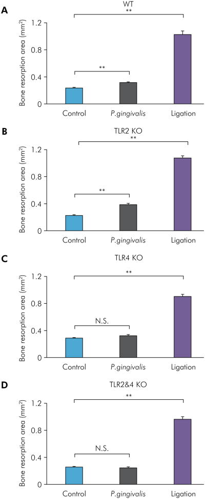
Figure 2.
Bone resorption analysis of P. gingivalis-induced and ligation-induced experimental periodontitis in WT, TLR2 KO, TLR4 KO, and TLR2&4 KO mice. The bone resorption area of P. gingivalis-induced and ligation-induced experimental periodontitis was measured and analyzed with ImageJ software on buccal and palatal surfaces in WT mice (A), TLR2 KO mice (B), TLR4 KO mice (C), and TLR2&4 KO mice (D) (means ± SE, n = 5 mice per group, **p < 0.01, N.S. = no significant difference). For each segment, a standard calibrator was used for calibration at the same magnification for all images.

