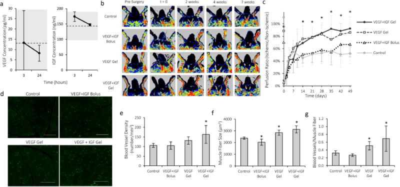
Figure 4.

Treatment with VEGF-containing alginate gels improves regional perfusion and angiogenesis in the ischemic rabbit hindlimb. a) Serum concentration of VEGF and IGF after intramuscular injection of gels into ischemic hind-limbs of rabbits. The dotted lines represent the average serum growth factor concentration measured prior to injection, and the grey regions mark one standard deviation for the baseline levels. b) Representative LDPI images of rabbit hindlimbs treated with VEGF+IGF alginate gels, VEGF gels, a bolus of VEGF and IGF (without alginate gel), or untreated control animals. c) Quantification of perfusion recovery after hindlimb ischemia surgery (n=6–9). *p < 0.05 compared to control condition for both VEGF and VEGF+IGF. d) Fluorescent images of capillaries stained for CD31 (green) in rabbit hind-limb muscle tissue. Scale bars 200μm. e) Capillary densities in rabbit hindlimb muscle seven weeks after surgery. f) Average muscle fiber size in treated region of rabbit hindlimb. g) Number of capillaries per muscle fiber in rabbit hindlimb. Values are Mean±SD. *p < 0.05 compared to control condition.
