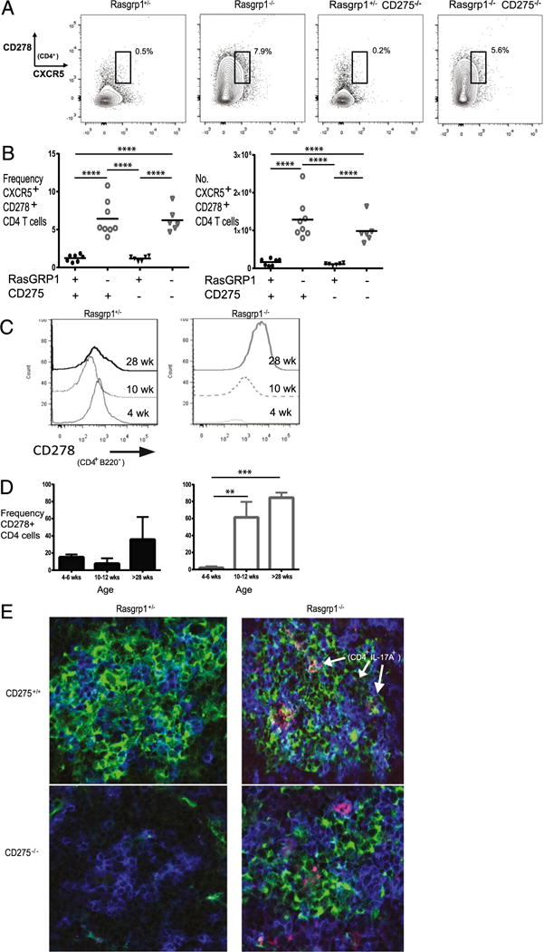
FIGURE 4.

CD278 levels and localization of IL-17–producing CD4 T cells in spleens of Rasgrp1−/− mice. (A) Frequency of CXCR5+ CD278+ CD4 T cells in representative Rasgrp1+/− littermate control and Rasgrp1−/− mice, as well as in both sets of mice lacking CD275. (B) Summary of the frequency (left panel) and number (right panel) of CXCR5+ CD278+ CD4 T cells for all groups of mice; each individual symbol represents the value for a single mouse (one-way ANOVA with Tukey’s multiple comparison test, p < 0.0001). (C) Representative graphs of CD278 levels on CD4 T cells from spleens of 4-, 10-, and 28-wk-old Rasgrp1+/− littermate control mice (left panel) and Rasgrp1−/− mice (right panel). (D) Summary of age-related changes from multiple mice (**p < 0.01, ***p < 0.001, t test). (E) Representative splenic sections with GCs from 12-wk-old littermate control mice (upper left panel), Rasgrp1−/− mice (upper right panel), CD275−/− mice (lower left panel), and Rasgrp1−/− CD275−/− mice (DKO, lower right) were examined by confocal microscopy for GCs (PNA, green), CD4 (blue), and IL-17 (red) (original magnification ×600). Note that no GC was detected in the section from the CD275−/− mouse.
