
Figure 1. The SE ligand.
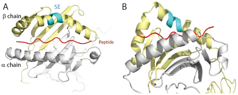
Crystal structure of HLA-DR4 (DRB1*04:01) molecule in a ‘top” (A) and ‘side’ (B) views. The DRα chain is colored in gray; the DRβ chain is shown in yellow and the groove peptide is shown in red. The SE (residues 70-74) is shown in cyan.