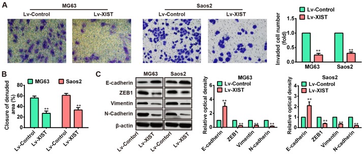
Figure 3.
Overexpression of XIST inhibited the migration and invasion of OS cells in vitro. (A) Invasion assay (use matrigel Transwell chambers) in MG63 and Saos2 cells was performed to determined cell invasiveness after infection with Lv-XIST (MOI=20). (B) Wound healing assay to evaluate the effect of XIST on cell migration in MG63 and Saos2 cells. (C) EMT-relatived proteins were detected by western blotting in MG63 and Saos2 cells, and Lv-XIST increased the expression levels of epithelial marker E-cadherin while reduced the level of mesenchymal markers such as ZEB1, vimentin and N-cadherin. **P<0.01 vs. Lv-control group. Data are presented as mean ± SD from three independent experiments.

