Figure 4.
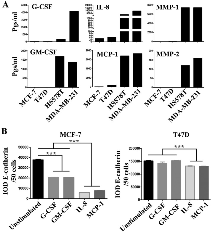
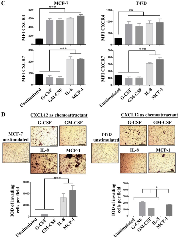
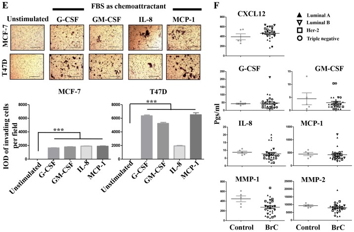
(A–D) The role of pro-inflammatory mediators in the inducible-invasive phenotype. (A) Milliplex assays were performed to determine the concentration of pro-inflammatory mediators and metalloproteinases (expressed in pgs/ml) in all the CMs; only analytes exhibiting significant differences between the CMs of NA- and HA-BrC cells are shown. MCF-7 and T47D cells were cultured with 100 ng/ml of any of the following: G-CSF, GM-CSF, IL-8 or MCP-1 for 72 h. (B) Analysis of the EMT marker E-cadherin by IF. (C) Plots of the analysis of CXCR4 and CXCR7 chemokine receptor expression by flow cytometry. Invasion assays using CXCL12 (D) as chemoattractant. Representative images and plots of resulting data are shown. Data represent the mean ± SEM from 3 independent experiments; *P<0.05, **P<0.01 and ***P<0.001. In the panels of T47D cells of (D), IL-8 was significantly different than the other cytokines (*P<0.05). Scale bars indicate 100 µm and magnification, ×100. (E and F) The role of pro-inflammatory mediators in the inducible-invasive phenotype. Invasion assays using fetal bovine serum (FBS) (E) as chemoattractant. (F) A Milliplex assay was performed to determine the sera concentration of the pro-inflammatory mediators and metalloproteinases of interest in BrC patients and controls. Representative images and plots of resulting data are shown. Data represent the mean ± SEM from 2 independent experiments; ***P<0.001. Only two duplicates were analyzed (F). Scale bars indicate 100 µm and magnification, ×100.


