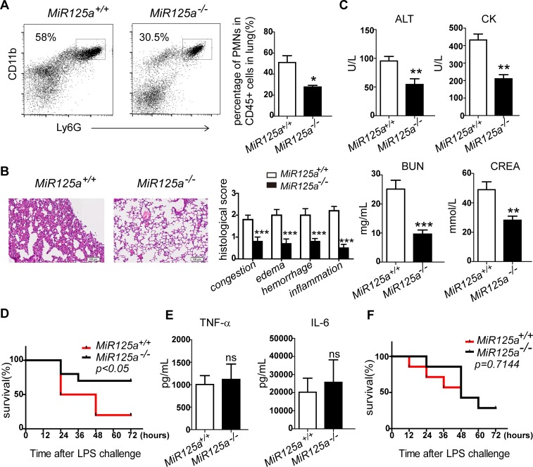
Fig 2. Lower mortality and neutrophil infiltration in LPS-induced lethal septic shock in MiR125a-/- mice.
(A) Flow cytometry analysis of infiltrating neutrophils from lungs of MiR125a+/+and MiR125a-/- mice challenged with 25 mg/kg LPS after 24 hours. Single cell suspensions of lung cells were previously gated with CD45. Neutrophils were stained with CD11b-Percp cy5.5 and Ly6G-APC. Bar graph shows the average percentage of infiltrating neutrophils (mean±s.d.,n = 3 mice of each genotype). (B) Hematoxylinand-eosin staining of lung sections from WT and KO mice 24 hours after 25 mg/kg LPS injection. Bar graph is the histopathological severity score of lung sections. Histopathological severity of randomly selected fields from the lung sections were graded as 0 (no findings or normal), 1 (mild), 2 (moderate) or 3 (severe) for each of the four parameters(congestion, edema, hemorrhage and inflammation). Theses results were confirmed by a blinded independent researcher. (C) Serum concentrations of aspartate aminotransferase (ALT), blood urea nitrogen (BUN), creatine kinase (CK) and creatinine (CREA) in MiR125a+/+and MiR125a-/- mice 24 h after injection of 25 mg/kg LPS (mean±s.d.,n = 5 mice of each genotype,). (D) Survival of MiR125a+/+and MiR125a-/- mice (n = 10 each genotype) intraperitoneally challenged with 45 mg/kg LPS. Data are presented as a Kaplan-Meier plot. P<0.05 (log-rank test). (E) TNF-α and IL-6 concentrations in serum of MiR125a+/+and MiR125a-/- mice 2h after intraperitoneal injection of 45 mg/kg LPS (mean±s.d., n = 5 mice of each genotype). ns, no significant difference (Student’s t-test). (F) Wild-type mice were first depleted of endogenous macrophages by pre-treatment with clodronate liposomes and then were transplanted with 1x10⁷ MiR125a+/+and MiR125a-/- bone marrow derived macrophages 6 hours before intraperitoneal injection with 45 mg/kg LPS. Survival percentage of these mice are presented as a Kaplan-Meier plot (n = 7 mice of each genotype;p = 0.7114, log-rank test).*P<0.05,**P<0.01, ***P<0.001.

