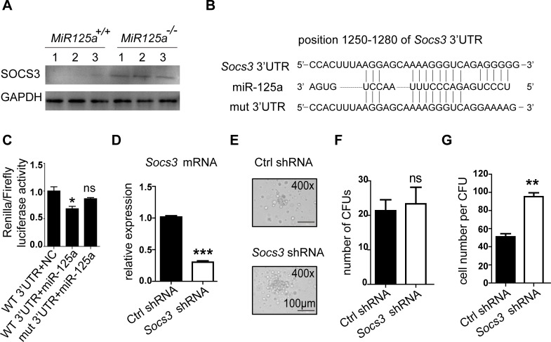
Fig 7. Socs3 ia a target of miR-125a.
(A) Protein expression of SOCS3 in bone marrow neutrophils from MiR125a+/+ and MiR125a-/- mice. Cell lysates were analyzed by immunoblot using SOCS3 antibody. (B) Schematic presentation of a potential miR-125a binding sites in the 3’UTR regions of Socs3. Sequences below indicate the mutant form of this site. (C) Luciferase reporter gene assay performed on 293T cells transfected with plasmids on which the luciferase reporter gene fused to the fragment of wild-type or mutant 3’UTRs of Socs3. Values were normalized to a firefly gene’s activity on the same construct (mean±s.d., n = 3). (D) The mRNA expression of Socs3 in sorted GFP+ GMPs which were transduced with retrovirus of Socs3 shRNA or a control(Ctrl) shRNA. (E-G) 1000 GMPs were sorted from MiR125a-/- bone marrow lin- cells which were transduced with retrovirus of Socs3 shRNA or a Ctrl shRNA and then cultivated in G-CSF containing methylcellulose media. Photographed CFUs (E), colony numbers (F) and cell number per CFUs (G) were shown. Representative data were from three independent experiments. **P<0.01 (Student’s t-test).

