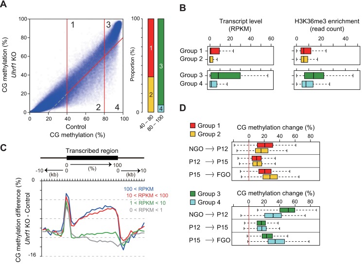
Fig 5. UHRF1 facilitates de novo methylation in inactive regions during late oocyte growth.
(A) The classification of the 10-kb genomic regions that were moderately (40–80%) or highly (≥80%) methylated in the control FGOs, based on the extent of methylation change (≥20% or not) in Uhrf1 KO FGOs (Groups 1–4). The right panel shows the proportion of moderately and highly methylated 10-kb regions belonging to each group. (B) The distribution of the transcript levels (RPKM) and H3K36me3 enrichment levels [20] (corrected read count) of the 10-kb regions belonging to each group. The bar in each box indicates the median value. (C) Differences in CG methylation across the transcribed regions between control and Uhrf1 KO FGOs. The transcribed regions were classified into four categories based on the expression levels (RPKM) in control FGOs and the results for the respective groups are shown separately. (D) The changes in CG methylation occurring in the three different stages of oocyte growth were determined for the 10-kb regions of the respective groups.

