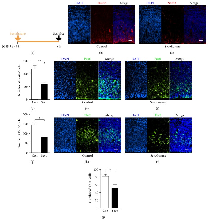
Figure 4.
Maternal sevoflurane exposure inhibited the expansion of neural progenitors in the fetal PFC. (a) Schematic diagram of the timing of sevoflurane exposure and sacrifice to assess the abundance of neural progenitors. (b, c) Nestin (red) immunofluorescence and DAPI staining (blue) in the cortical plate at G15.5. Scale bars, 20 μm. (d) Quantification of the nestin+ cells of the control and sevoflurane groups. (e, f) Pax6 (green) immunofluorescence and DAPI staining (blue) in the cortical plate at G15.5. Scale bars, 20 μm. (g) Quantification of the Pax6+ cells of the control and sevoflurane groups. (h, i) Tbr2 (green) immunofluorescence and DAPI staining (blue) in the cortical plate at G15.5. Scale bars, 20 μm. (j) Quantification of the Tbr2+ cells of the control and sevoflurane groups. Data are expressed as the mean ± SEM. ∗P < 0.05, ∗∗P < 0.01, and ∗∗∗P < 0.001.

