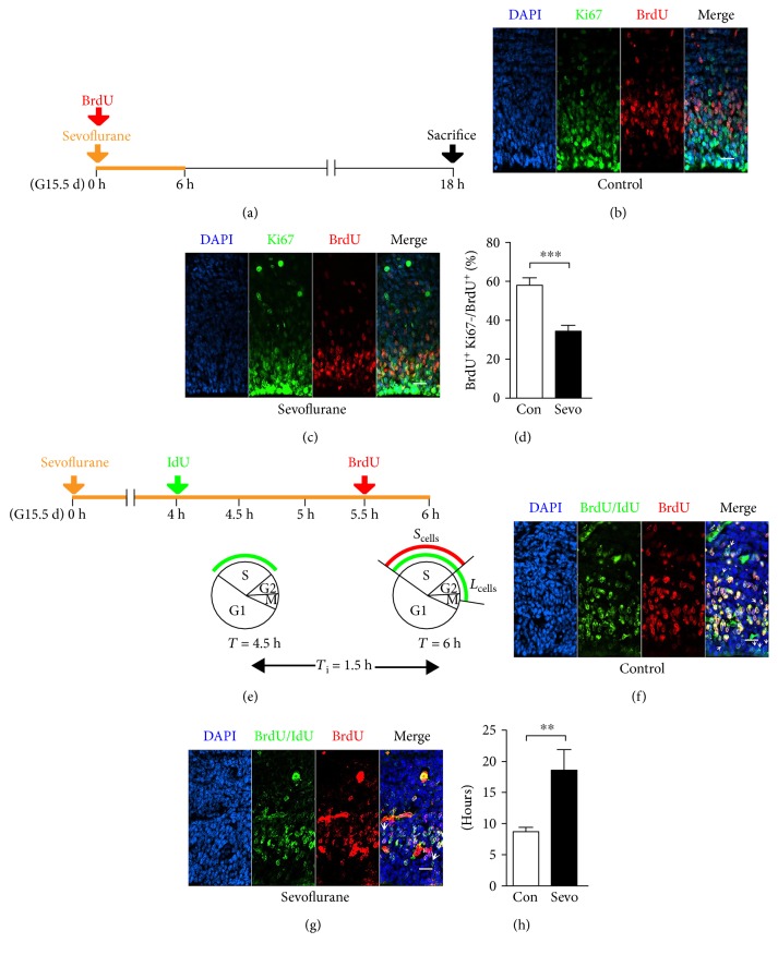
Figure 5.
Maternal sevoflurane exposure decreased cell cycle exit and increased S-phase duration of neural progenitors in the fetal PFC. (a) Schematic diagram of the timing of sevoflurane exposure, BrdU injection, and sacrifice to assess the proportion of the cell cycle exit. (b, c) Coronal sections of the PFC from G16.5 mice were immunostained for BrdU (red) and Ki67 (green) at G16.5. Scale bars, 20 μm. (d) Numbers of BrdU+Ki67− cells are expressed as the numbers of BrdU+ cells. (e) Schematic diagram of the timing of sevoflurane exposure, IdU injection, and BrdU administration to assess the S-phase duration. Scells = cells labeled with BrdU; Lcells = cells labeled with IdU but not BrdU. (f, g) Coronal section through the cortex of the G15.5 fetal brain immunostained with antibodies specific for both BrdU and IdU (green) and BrdU alone (red) to identify Lcells (green-only cells) and Scells (red and green double-labeled cells). Arrowheads indicate Lcells. Scale bars, 20 μm. (h) Quantification of the length of S-phase in G15.5 embryos. Data are expressed as the mean ± SEM. ∗∗P < 0.01 and ∗∗∗P < 0.001.

