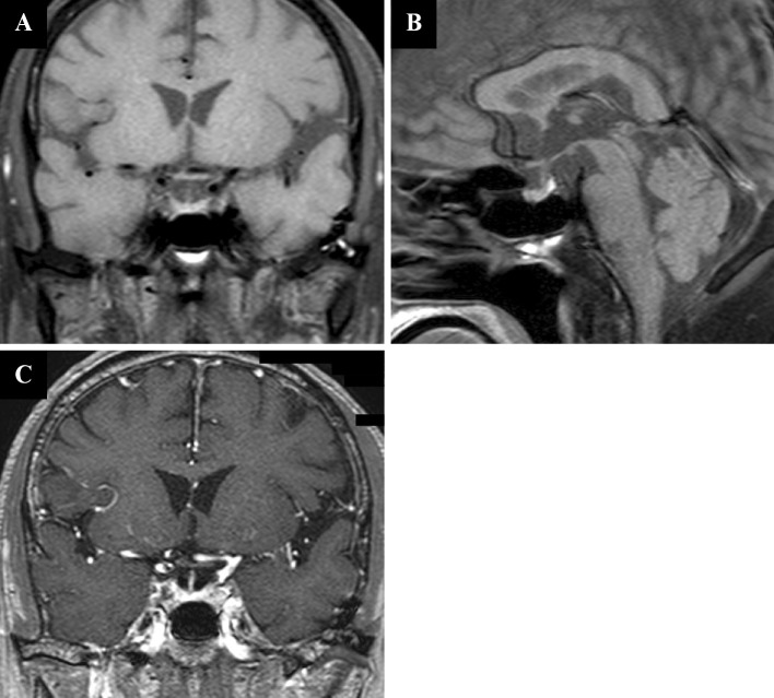
Figure 1.
A: Coronal section of a T1-weighed magnetic resonance image (MRI) scan of the brain reveals no enlargement of the pituitary gland or the stalk’s thickness. B: Sagittal section of a T1-weighed brain MRI reveals a normal signal at the posterior lobe of the pituitary gland. C: Coronal section of a contrast-enhanced T1-weighed MRI scan of the brain reveals no abnormality in the pituitary gland.

