Figure.
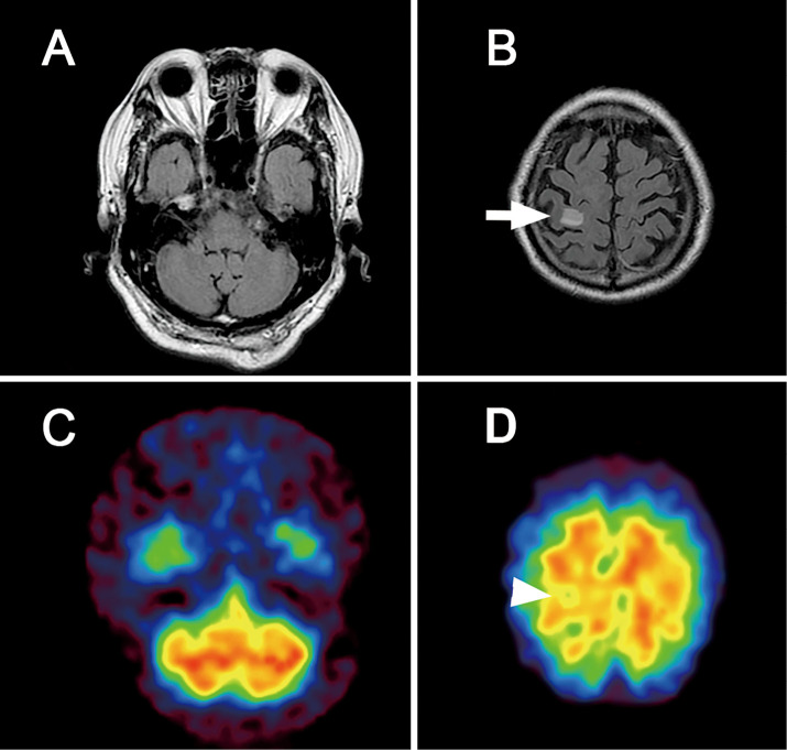
(A, B) Brain fluid attenuated inversion recovery (FLAIR) and single photon emission computed tomography (SPECT) images from a patient with ataxic hemiparesis. FLAIR imaging revealed no abnormalities in the cerebellum or brainstem that were associated with the lesions of the postcentral gyrus (white arrow). (C, D) Brain SPECT revealed a slight decrease in the cerebral blood flow around the postcentral gyrus (white arrowheads); however, no signs of crossed cerebellar diaschisis were observed.

