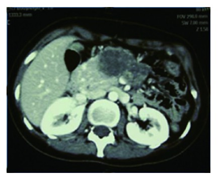
Figure 1.

Preoperative computed tomography view of a 26-year-old female patient with a diagnosis of pseudo-papillary tumors. The figure shows that the lesion occupied the body and neck region with necrosis of the tail on preoperative computed tomography imaging. Normal pancreatic tissue is observed in the ventral pancreas region.
