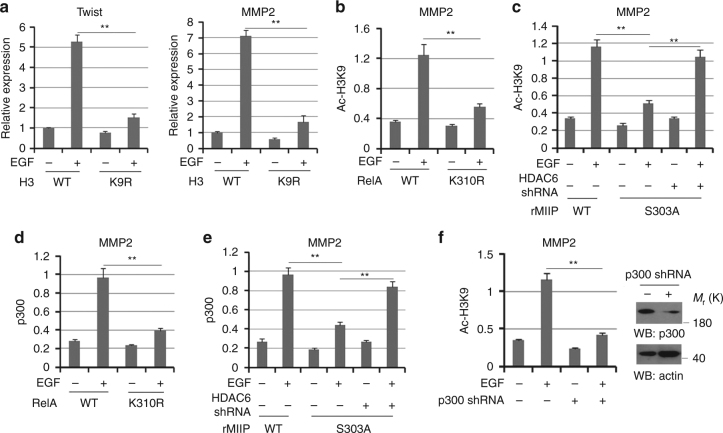
Fig. 5.
MIIP–RelA facilitates H3-K9 acetylation at promoter region. a HCT116 cells expressed with WT H3 or H3 K9R were treated with or without EGF (100 ng/ml) for 10 h. Relative mRNA levels were analyzed by q-PCR. b, d HCT116 cells expressed with WT RelA or RelA K310R were treated with or without EGF for 10 h (100 ng/ml). c, e HCT116 cells expressed with WT MIIP or MIIP S303A were overexpressed with or without HDAC6; cells were treated with or without EGF (100 ng/ml) for 10 h. f HCT116 cells transfected with or without plasmid for expressing p300 shRNA were treated with or without EGF (100 ng/ml) for 10 h. In b–f, ChIP analyses with indicated antibodies were performed. The primers covering RelA binding site of MMP2 gene promoter region were used for the q-PCR. The Y axis shows the value normalized to the input. In a–f, the values are presented as mean ± s.e.m. (n = 3 independent experiments), ** represents P < 0.01 (Student’s t-test) between the indicated groups

