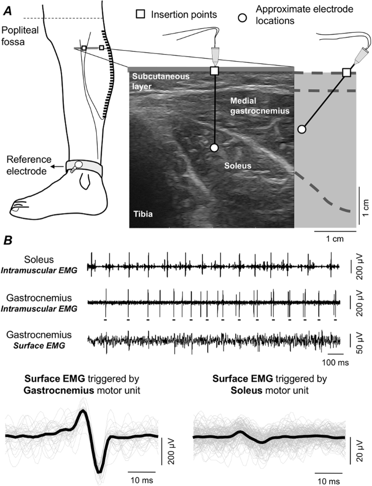
Figure 2.
Surface and intramuscular EMG detection and analysis. (A) schematic illustration of the positioning of intramuscular and surface electrodes. Almost the whole gastrocnemius muscle was covered proximo-distally by 32 surface electrodes. With the guidance of ultrasound scanning (right panel), wire electrodes were inserted into gastrocnemius and soleus, roughly beneath the parasagittal section defined by the surface electrodes. (B) examples of intramuscular EMGs collected from both muscles and a single, differential surface recording (5 mm inter-electrode distance) are shown. Grey traces in the bottom left correspond to short (60 ms) epochs of surface EMGs triggered with the firing instants of a motor unit identified from the gastrocnemius, intramuscular EMG (cf. short horizontal bars underneath large spikes). These epochs were then averaged, producing the spike, triggered average representation of the motor unit action potential in the surface EMG (black trace; cf. Methods). An example of the surface representation of a motor unit from soleus is shown in the right bottom panel.

