Figure 4.
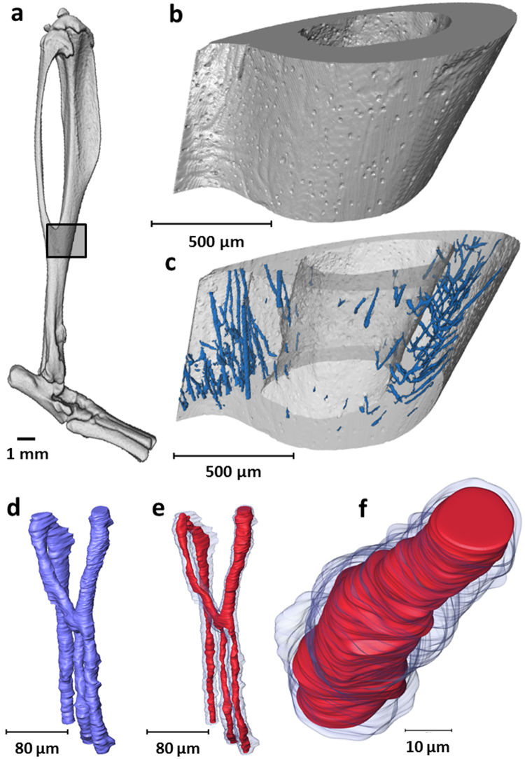
3D visualisation and extraction of intracortical blood vessel. (a) 3D rendering of murine tibia with identification of the tibiofibular junction. (b) 3D rendering of scanned tibiofibular junction region (1.3 µm voxel size) and (c) detection of intracortical canals (blue) as a negative imprint of the mineralised tissue (extracted from 1.3 µm voxel size dataset). (d) Magnified area of the 3D intracortical network (extracted from 0.325 µm voxel size dataset) and (e) detection of the soft tissue comprising blood vessels (red) within intracortical canals (blue) (extracted from 0.325 µm voxel size dataset). (f) Magnified segment of blood vessel (red) within intracortical canal (blue) (extracted from 0.325 µm voxel size dataset).

