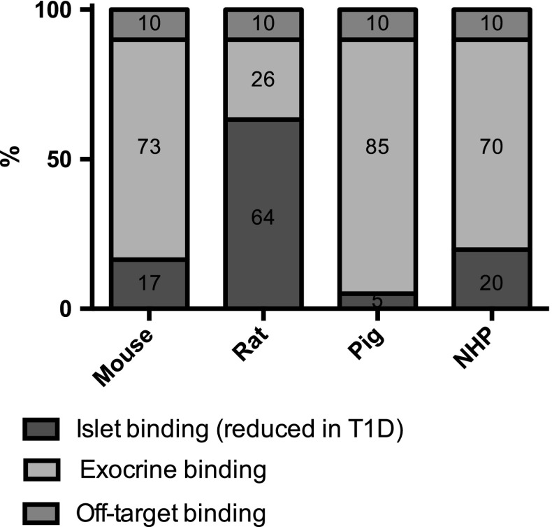
Fig. 5.
Theoretical binding pattern of [68Ga]Ga-DO3A-VS-Cys40-Exendin4 in pancreas in vivo, based on the species dependent islet contrast observed by ex vivo autoradiography. Each entire bar represents 100% of the uptake in pancreas in respective species, and each colored field is proportional in area to its predicted contribution to the total in vivo PET signal. The contribution of GLP-1R mediated binding in islets is indicated in blue. The predicted GLP-1R mediated binding in exocrine pancreas is indicated in bright red. Off-target binding, i.e., non-specific binding not mediated by GLP-1R throughout the pancreas, is indicated in yellow (color figure online)

