Figure 1.
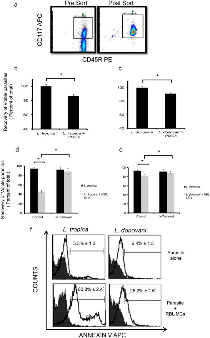
Cell death of promastigotes on co-culture with MCs. PMCs were isolated from Peritoneal lavage of female BALB/c mice and were sorted through flowcytometer (Fig. 1a). PMCs were co-cultured with L. tropica and L. donovani in 96 well plate for 24 h at MOI (1:10). MTT assay was done. The Y-axis represents the relative amount of viable cells after normalization to the Leishmania control (Fig. 1b and c). 0.1 × 10⁶ MCs were seeded in 48 well cell culture plate and cultured overnight in CO2 incubator. L. donovani and L. tropica were added at MOI 1:10. After indicated time points parasites were removed cell viability was counted using trypan blue exclusion method n = 3. Panel d and e represent % cell viability of L. tropica and L. donovani on co-culture with MCs as well as % cell viability of L. tropica and L. donovani on co-culture with MCs in transwell system respectively. 0.1 million cells were seeded in 48 well cell culture plate. Leishmania were syringe separated and added at MOI 1:10 for 24 h. Parasites were harvested washed followed by adding 50 μl of Annexin Binding Buffer and were processed as mentioned in materials and methods. Representative histograms (solid black line) in panels show Annexin APC positive cells in comparison to unstained (filled grey)(Fig. 1f). Each point represents mean ± SEM of values obtained from three independent assays.

